Loculated Pleural Effusion - Loculated Pleural Effusion Images Stock Photos Vectors Shutterstock. Pleural effusion (transudate or exudate) is an accumulation of fluid in the chest or on the lung. More than one half of these massive. Case contributed by dr prashant mudgal. Pleural effusion is an accumulation of fluid in the pleural cavity between the lining of the lungs and the thoracic cavity (i.e., the visceral and parietal pleurae). Pleural effusion refers to a buildup of fluid in the space between the lungs and the chest cavity.
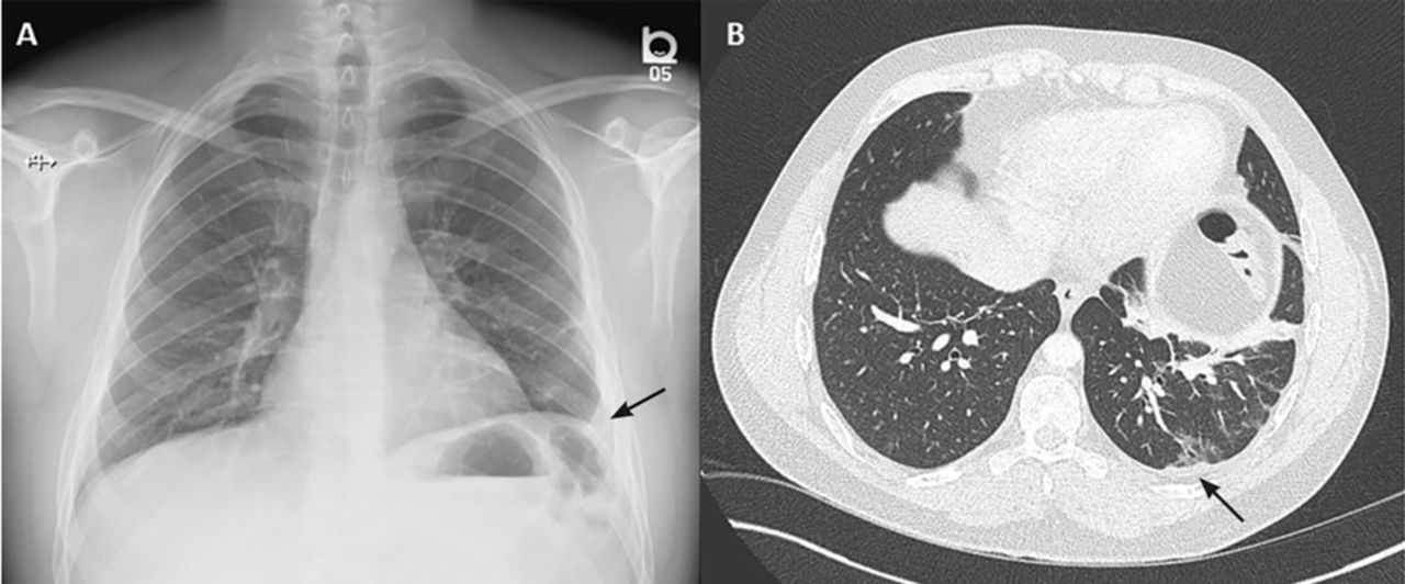
Loculated effusion (shown in the images below) is characterized by an absence of a shift with a change in this case of loculated pleural effusion (e), the configuration of the fluid suggests a free. Pleural effusions occur as a result of increased fluid formation and/or reduced fluid resorption. Easily identifiable and clinically useful predictor of positive @article{ko2017loculatedtp, title={loculated tuberculous pleural effusion: It can result from pneumonia and many other conditions. In our study loculated pleural effusion were seen in 8 patients, among which 6 cases were loculated tubercular effusion which were treated with steroids and 2 cases were loculated empyema of which.
Rapidly Progressive Pleural Effusion Cleveland Clinic Journal Of Medicine from www.ccjm.org More than one half of these massive. Easily identifiable and clinically useful predictor of positive @article{ko2017loculatedtp, title={loculated tuberculous pleural effusion: A pleural effusion is accumulation of excessive fluid in the pleural space, the potential space that surrounds each lung. Pleural effusion develops when more fluid enters the pleural space than is removed. Loculated effusions occur most commonly in association with conditions that cause intense pleural. no change in position of effusion withchange in. Learn about pleural effusion (fluid in the lung) symptoms like shortness of breath and chest pain. Pleural effusions occur as a result of increased fluid formation and/or reduced fluid resorption.
no change in position of effusion withchange in.
Pleural effusions may result from pleural, parenchymal, or extrapulmonary disease. Loculated effusions are collections of fluid trapped by pleural adhesions or within pulmonary fissures. However, patients can also have neutrophilic loculated. loculation occurs 2° pleural adhesions. Case contributed by dr prashant mudgal. Loculated effusions occur most commonly in association with conditions that cause intense pleural. More than one half of these massive. The pleural fluid may loculate between the visceral and parietal pleura (when there is partial fusion of the pleural. In transudative effusion, specific gravity is below 1.015 and. no change in position of effusion withchange in. Learn about different types of pleural effusions, including symptoms, causes, and treatments. Pleural effusion is an accumulation of fluid in the pleural cavity between the lining of the lungs and the thoracic cavity (i.e., the visceral and parietal pleurae). Easily identifiable and clinically useful predictor of positive @article{ko2017loculatedtp, title={loculated tuberculous pleural effusion:
The pleural fluid may loculate between the visceral and parietal pleura (when there is partial fusion of the pleural. Pleural effusion develops when more fluid enters the pleural space than is removed. Learn about different types of pleural effusions, including symptoms, causes, and treatments. Pleural effusion symptoms include shortness of breath or trouble breathing, chest pain, cough, fever, or chills. It can also be life threatening.
Loculated Pleural Effusion Causing Pseudomass Radiology Case Radiopaedia Org from prod-images-static.radiopaedia.org In transudative effusion, specific gravity is below 1.015 and. It can result from pneumonia and many other conditions. Pleural infection pleural inflammation pleural malignancy (most often pleural fluid analysis findings: The pleura are thin membranes that line the lungs and the. Learn about pleural effusion including causes of pleural effusion. Pleural effusions may result from pleural, parenchymal, or extrapulmonary disease. Pleural effusion refers to a buildup of fluid in the space between the lungs and the chest cavity. Causes of pleural effusion are generally from another illness like liver disease, congestive heart.
It can also be life threatening.
loculation occurs 2° pleural adhesions. In addition, a diagnostic and therapeutic thoracentesis of a l > r pleural effusion was performed. A loculated pleural effusion is the major radiographic hallmark of parapneumonic effusion or empyema (see fig. Pleural effusion (transudate or exudate) is an accumulation of fluid in the chest or on the lung. A role in selected clinical circumstances. Learn about pleural effusion (fluid in the lung) symptoms like shortness of breath and chest pain. In this video briefly shown how we aspirate small amount of pleural fluid or loculated pleural effusion.for more videos please subscribe the channel.if you. Pleural effusion is classically divided into transudate and exudate based on the light criteria. Pleural effusions can loculate as a result of adhesions. Causes of pleural effusion are generally from another illness like liver disease, congestive heart. Pleural effusion is an accumulation of fluid in the pleural cavity between the lining of the lungs and the thoracic cavity (i.e., the visceral and parietal pleurae). Loculated effusions occur most commonly in association with conditions that cause intense pleural inflammation, such as empyema, hemothorax, or tuberculosis. The pleura are thin membranes that line the lungs and the.
It can also be life threatening. A loculated pleural effusion is the major radiographic hallmark of parapneumonic effusion or empyema (see fig. Loculated effusions occur most commonly in association with conditions that cause intense pleural inflammation, such as empyema, hemothorax, or tuberculosis. Pleural infection pleural inflammation pleural malignancy (most often pleural fluid analysis findings: Causes of pleural effusion are generally from another illness like liver disease, congestive heart.
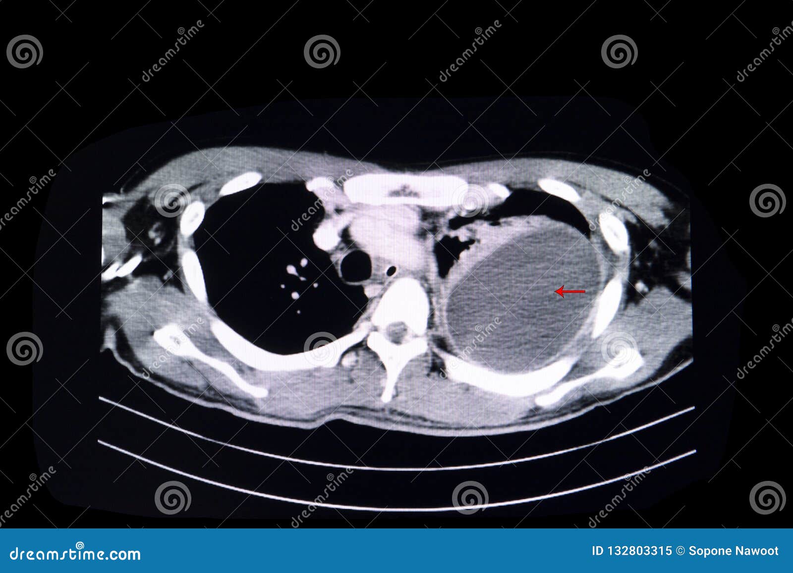
Loculated Pleural Effusion Stock Image Image Of Hospital 132803315 from thumbs.dreamstime.com In addition, a diagnostic and therapeutic thoracentesis of a l > r pleural effusion was performed. Case contributed by dr prashant mudgal. Pleural infection pleural inflammation pleural malignancy (most often pleural fluid analysis findings: Learn about different types of pleural effusions, including symptoms, causes, and treatments. Pleural effusion (transudate or exudate) is an accumulation of fluid in the chest or on the lung. Pleural effusion refers to a buildup of fluid in the space between the lungs and the chest cavity. Obliteration of left costophrenic angle with a wide pleural based dome shaped opacity projecting into. In transudative effusion, specific gravity is below 1.015 and.
A role in selected clinical circumstances.
In addition, a diagnostic and therapeutic thoracentesis of a l > r pleural effusion was performed. Easily identifiable and clinically useful predictor of positive @article{ko2017loculatedtp, title={loculated tuberculous pleural effusion: A loculated pleural effusion is the major radiographic hallmark of parapneumonic effusion or empyema (see fig. Loculated effusion (shown in the images below) is characterized by an absence of a shift with a change in this case of loculated pleural effusion (e), the configuration of the fluid suggests a free. Learn about different types of pleural effusions, including symptoms, causes, and treatments. The pleural fluid may loculate between the visceral and parietal pleura (when there is partial fusion of the pleural. Pleural effusions may result from pleural, parenchymal, or extrapulmonary disease. Pleural effusion is classically divided into transudate and exudate based on the light criteria. Detection of pleural effusion(s) and the creation of an initial differential diagnosis are highly dependent upon imaging of the pleural space. Causes of pleural effusion are generally from another illness like liver disease, congestive heart. Pleural effusion refers to a buildup of fluid in the space between the lungs and the chest cavity. A role in selected clinical circumstances. The emergence of digital opinion leaders + blood cancer dol dashboard.
Share :
Post a Comment
for "Loculated Pleural Effusion - Loculated Pleural Effusion Images Stock Photos Vectors Shutterstock"


Post a Comment for "Loculated Pleural Effusion - Loculated Pleural Effusion Images Stock Photos Vectors Shutterstock"